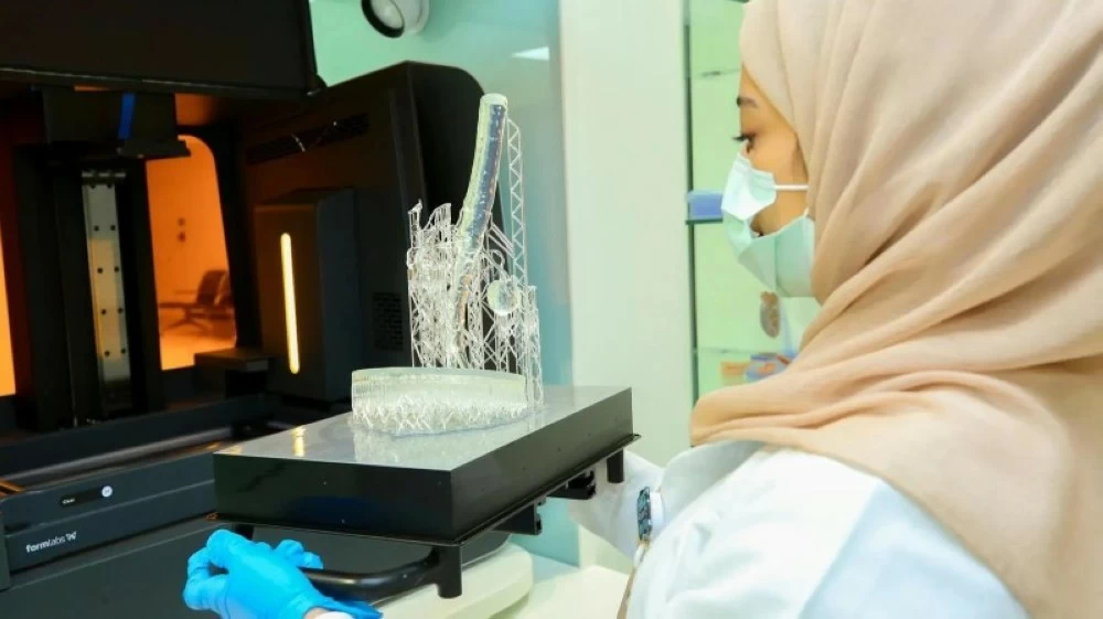
نجح فريق طبي بالسعودية في إجراء جراحة دقيقة لاستئصال ورم معقّد يقع في أعلى عظم الفخذ ويمتد إلى مفصل الورك الأيسر، وذلك باستخدام تقنية الطباعة ثلاثية الأبعاد التي أسهمت في تصميم قوالب عظمية مخصصة مكنت الجراحين من إزالة الورم بالكامل مع المحافظة على الطرف وتمكين المريض من المشي مباشرة بعد العملية.
واعتمدت العملية على قوالب ثلاثية الأبعاد عالية الدقة استُخرجت بناءً على تصوير إشعاعي عالي الدقة، ما أتاح استئصال الورم بهوامش آمنة وتقليل فقدان العظم إلى الحد الأدنى، قبل تركيب مفصل صناعي متطور ساعد المريض على استعادة الحركة بشكل سريع وآمن.
وجرى تنفيذ هذه الجراحة المتقدمة في مستشفى الملك فيصل التخصصي ومركز الأبحاث، إذ تفتح هذه التقنية آفاقًا واسعة لتطبيقها على مرضى يعانون من أورام في مناطق معقّدة من العظام، بما يسهم في تقليص مدة العمليات الجراحية، وتمكين المرضى من التعافي بشكل أسرع والعودة إلى ممارسة حياتهم الطبيعية.
A medical team in Saudi Arabia successfully performed a precise surgery to remove a complex tumor located at the top of the femur, extending to the left hip joint, using 3D printing technology that contributed to the design of customized bone templates. This enabled the surgeons to completely remove the tumor while preserving the limb and allowing the patient to walk immediately after the operation.
The procedure relied on high-precision 3D templates extracted based on high-resolution imaging, which allowed for the tumor to be removed with safe margins and minimized bone loss, before installing an advanced artificial joint that helped the patient regain mobility quickly and safely.
This advanced surgery was performed at King Faisal Specialist Hospital and Research Center, as this technology opens wide horizons for its application on patients suffering from tumors in complex areas of the bones, contributing to reducing the duration of surgical operations and enabling patients to recover faster and return to their normal lives.