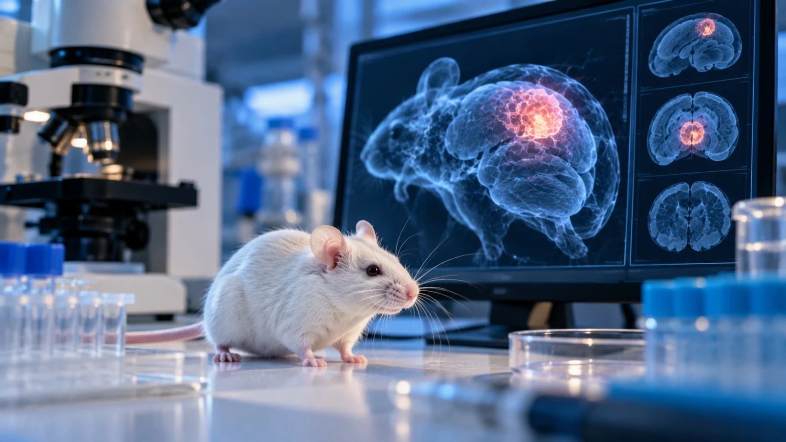
نجح باحثون من جامعة ستانفورد الأمريكية في زراعة أنسجة بشرية شبيهة بالقشرة الدماغية داخل فئران معدلة وراثياً، في تجربة أتاحت للأنسجة النمو وتكوين اتصالات وظيفية داخل الجهاز العصبي للحيوان، بهدف تطوير نموذج جديد لدراسة نمو الدماغ البشري واضطراباته.
ونُشرت نتائج الدراسة في دورية Nature العلمية، واعتمد الباحثون على «عضيات قشرية» بشرية، وهي أنسجة ثلاثية الأبعاد نُميت في المختبر من خلايا بشرية أُعيدت برمجتها إلى خلايا جذعية، قبل تطويرها إلى أنسجة تحاكي جوانب من القشرة الدماغية البشرية.
مساحة للنمو
وعدّل باحثو ستانفورد الفئران وراثياً بحيث تفتقر إلى معظم القشرة الدماغية والحُصين، ما وفر مساحة للأنسجة البشرية المزروعة كي تنمو وتتطور.
وزُرعت العضيات القشرية البشرية في الفئران بعد ولادتها بفترة قصيرة، وأظهرت النتائج أن الأنسجة نمت وأنتجت أنواعاً متعددة من الخلايا القشرية، كما أقامت الخلايا البشرية اتصالات مع خلايا دماغ الفئران والحبل الشوكي.
ليست «أدمغة بشرية»
ولا تعني التجربة أن الفئران أصبحت تمتلك أدمغة بشرية، إذ ظلت الأنسجة المزروعة غير ناضجة ولم تكن منظمة أو متصلة بالطريقة نفسها الموجودة في الدماغ البشري.
وأطلق الباحثون على النموذج اسم «الفئران ذات القشرة الغريبة» (Xenocortical mice)، في إشارة إلى وجود أنسجة قشرية بشرية مزروعة داخل أدمغتها.
5 ساعات من نقص الأكسجين
ولاختبار إمكان استخدام النموذج في دراسة إصابات الدماغ، عرّض الباحثون بعض الفئران لخمس ساعات من انخفاض الأكسجين، في محاكاة لحالة يمكن أن تسهم في حدوث الشلل الدماغي لدى البشر عندما تقع أثناء الحمل أو قرب الولادة.
وأظهرت الفئران بعد تعرضها لنقص الأكسجين صعوبة أكبر في الحفاظ على التوازن والمشي، فيما كشفت التجربة حساسية الخلايا العصبية البشرية لنقص الأكسجين.
دراسة اضطرابات الدماغ
ويرى الباحثون أن النموذج الجديد يمكن أن يساعد على دراسة تطور أنسجة الدماغ البشري داخل كائن حي، وفهم الآليات المرتبطة باضطرابات عصبية ونفسية.
وقد يتيح النموذج دراسة حالات من بينها الفصام والصرع والشلل الدماغي وبعض أشكال التوحّد الشديدة، إضافة إلى اختبار تدخلات محتملة لفهم هذه الاضطرابات وعلاجها.
وتثير زراعة الأنسجة العصبية البشرية داخل الحيوانات اعتبارات أخلاقية تتعلق بالتجارب على الحيوانات وتأثير الأنسجة البشرية المزروعة، ما يستدعي استمرار الرقابة والضوابط الأخلاقية مع تطور هذا المجال البحثي.
Researchers from Stanford University have succeeded in growing human tissue resembling the cerebral cortex inside genetically modified mice, in an experiment that allowed the tissues to grow and form functional connections within the animal's nervous system, with the aim of developing a new model for studying human brain development and its disorders.
The results of the study were published in the scientific journal Nature, and the researchers relied on "cortical organoids," which are three-dimensional tissues grown in the lab from human cells that have been reprogrammed into stem cells, before developing them into tissues that mimic aspects of the human cerebral cortex.
Room for Growth
The Stanford researchers genetically modified the mice to lack most of the cerebral cortex and hippocampus, providing space for the implanted human tissues to grow and develop.
The human cortical organoids were implanted in the mice shortly after birth, and the results showed that the tissues grew and produced multiple types of cortical cells, while the human cells established connections with the mouse brain and spinal cord cells.
Not "Human Brains"
This experiment does not mean that the mice now have human brains, as the implanted tissues remained immature and were not organized or connected in the same way as found in the human brain.
The researchers named the model "xenocortical mice," referring to the presence of implanted human cortical tissues within their brains.
5 Hours of Oxygen Deprivation
To test the potential use of the model in studying brain injuries, the researchers exposed some of the mice to five hours of reduced oxygen, simulating a condition that can contribute to cerebral palsy in humans when it occurs during pregnancy or near birth.
The mice showed greater difficulty in maintaining balance and walking after being subjected to oxygen deprivation, while the experiment revealed the sensitivity of human neurons to low oxygen levels.
Studying Brain Disorders
The researchers believe that the new model could help study the development of human brain tissues within a living organism and understand the mechanisms associated with neurological and psychological disorders.
The model may allow for the study of conditions including schizophrenia, epilepsy, cerebral palsy, and some severe forms of autism, in addition to testing potential interventions to understand and treat these disorders.
The implantation of human neural tissues into animals raises ethical considerations related to animal testing and the impact of the implanted human tissues, necessitating ongoing oversight and ethical guidelines as this research field evolves.