


طور علماء في جامعة «سيتشينوف» الروسية وحدات بناء خلوية تمهد للطباعة الحيوية ثلاثية الأبعاد لأنسجة دواعم الأسنان، وهي منظومة الأنسجة المحيطة بالسن والمسؤولة عن تثبيته داخل الفك.
جمع الفريق العلمي بين خلايا اللثة البشرية وخلايا لب الأسنان، مما أسفر عن إنتاج هياكل أكثر استقراراً وتنظيماً وتوازناً من الناحية الميكانيكية، مقارنة بالوحدات التي تعتمد على نوع واحد من الخلايا.
طور العلماء عدة أنواع من الوحدات الخلوية ثلاثية الأبعاد، بعضها يحتوي على خلايا اللثة فقط، وبعضها على خلايا لب الأسنان فقط، بينما جمع بعضها الآخر بين النوعين، ثم قارنوا سلوك الوحدات المختلفة وكفاءة تنظيمها ومدى ملاءمتها لبناء هياكل نسيجية أكبر.
أظهرت النتائج أن الوحدات التي تجمع بين نوعي الخلايا كانت الأكثر نجاحاً؛ إذ أظهرت قدرة أكبر على التنظيم الذاتي وخصائص ميكانيكية أكثر توازناً، بينما كانت خلايا لب الأسنان أكثر ملاءمة لتكوين تراكيب كثيفة وقوية، وأسهمت خلايا اللثة بصورة أكبر في ترميم الأنسجة الرخوة والتئامها.
قالت بولينا بيكمولينا، مديرة مركز تصميم المصانع الحيوية: «إن الدراسة أظهرت قدرة الوحدات الخلوية المنظمة على محاكاة الطبيعة متعددة المكونات لأنسجة دواعم الأسنان، لتشكل أساساً لمزيد من أعمال الطباعة الحيوية لأنسجة مماثلة».
من المقرر أن يدمج الفريق العلمي هذه الوحدات في هياكل نسيجية كاملة، يمكن مستقبلاً تصميمها بصورة مخصصة لكل مريض، وفقاً لشكل الضرر وتشريح المنطقة المتضررة.
علماء روس يطورون وحدات خلوية تمهد للطباعة الحيوية ثلاثية الأبعاد لأنسجة دواعم الأسنان
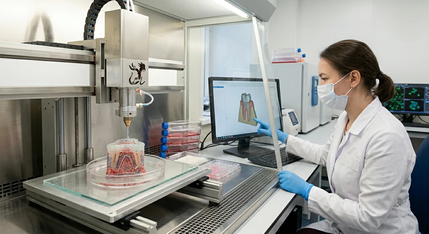


Scientists at the Russian "Sechenov" University have developed cellular building blocks that pave the way for 3D bioprinting of periodontal tissues, which are the tissues surrounding the tooth responsible for anchoring it within the jaw.
The research team combined human gum cells with dental pulp cells, resulting in the production of structures that are more stable, organized, and mechanically balanced compared to units relying on a single type of cell.
The scientists developed several types of 3D cellular units, some containing only gum cells, some containing only dental pulp cells, while others combined both types, and then compared the behavior of the different units, their organizational efficiency, and their suitability for building larger tissue structures.
The results showed that the units combining both cell types were the most successful; they exhibited greater self-organization capabilities and more balanced mechanical properties, while dental pulp cells were more suitable for forming dense and strong structures, and gum cells contributed more significantly to the repair and healing of soft tissues.
Polina Bikmullina, director of the Bioprinting Design Center, stated: "The study demonstrated the ability of organized cellular units to mimic the multi-component nature of periodontal tissues, forming a basis for further bioprinting work for similar tissues."
The research team is set to integrate these units into complete tissue structures, which can be custom-designed for each patient in the future, according to the shape of the damage and the anatomy of the affected area.


